Ovarian volume is more useful than taking a single measurement such as length. Why?
The ovary is a dynamic organ, and its size and shape change with age and during different phases of the menstrual cycle.
If you only measure one dimension, such as a length of 4 cm, it does not provide the full picture. You cannot tell whether the ovary is thin and flat or enlarged and bulky.
Because of this, a single measurement alone does not accurately describe the true size of the ovary.
Ovarian volume gives a better representation of the overall size. For this reason, measuring volume is considered the standard approach during ultrasound evaluation.
After reading this article, you will
- learn to measure the ovarian volume using standard protocol
- know the normal range of ovarian volume in different age group
- understand the common cause of enlarged ovary
Standard Technique to Measure Ovarian Volume
Transvaginal sonography (TVS) is considered the gold standard for measuring ovarian volume.
It is the most accurate method because it provides better resolution and detail of pelvic structures.
In daily clinical practice, both transabdominal and transvaginal ultrasound approaches are used.
However, TVS generally provides clearer visualization of the ovaries.
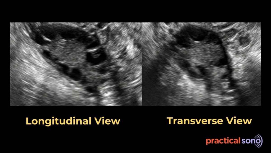
The first step is to identify the ovary and optimize the image before taking measurements.
Locating the Ovary
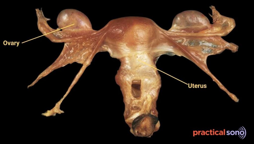
The ovary is usually located in the adnexal region. It typically lies:
- Lateral and posterior to the uterus
- Medial and anterior to the internal iliac vessels
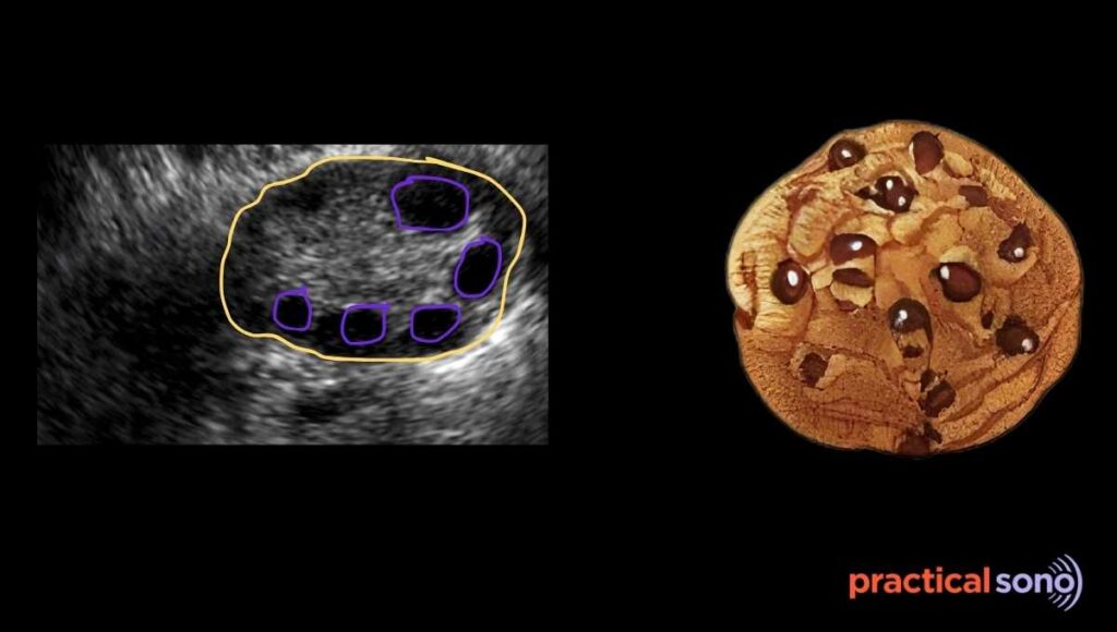
The ovary is almond-shaped and usually appears hypoechoic, meaning it looks darker than the surrounding pelvic fat and myometrium.
A typical ovary often shows multiple small follicles that create an appearance similar to “chocolate chips.”
When searching for the ovary, look for a hypoechoic oval structure with this chocolate chip appearance in the adnexal region, usually just lateral to the uterine fundus.
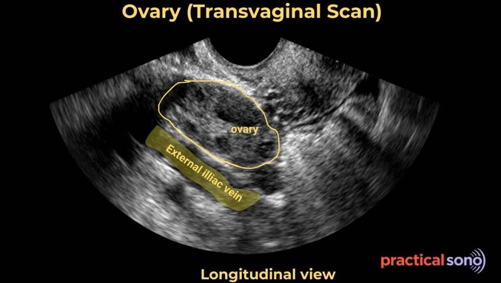
Sometimes, you may see an anechoic tubular structure in the adnexal region. This may represent the external iliac vein.
To confirm, apply color Doppler, which will demonstrate blood flow within the vessel.
The ovary is typically located just medial to these vessels, so carefully examine this area when searching for it.
Optimizing the Image
Once the ovary is identified, the image must be optimized before measurement. This step is essential for obtaining accurate dimensions.
Adjust the depth and focal zone so that the ovary occupies at least half of the ultrasound screen. This improves visualization and helps place the calipers accurately.
Turn on tissue harmonic imaging to improve border definition and make the ovarian margins clearer.
Reposition the probe if necessary to minimize bowel gas or acoustic shadowing, which can obscure the ovary.
Next, adjust the probe to obtain a true longitudinal view of the ovary. Ensure that the entire ovarian margin is clearly visible.
Only after achieving a clear and well-optimized image should you freeze the screen and begin measurements.
Measuring the Ovarian Dimensions
After obtaining the ideal image, press the Freeze button on the ultrasound machine. Then select the Measure or Calc option.
From the on-screen menu, choose Gynecology, followed by Ovary Volume.
To calculate ovarian volume, three dimensions must be measured:
- Length
- Depth (anteroposterior diameter)
- Width
Measuring Length and Depth:
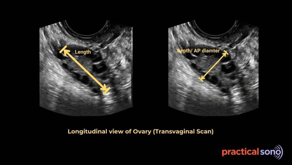
Length and depth are measured in the longitudinal view of the ovary.
First, measure the length. Place the first caliper on the superior (upper) margin of the ovary and the second caliper on the inferior (lower) margin. Always place the calipers on the outer border of the ovary.
Next, measure the depth, also called the anteroposterior diameter. In the same longitudinal view, place one caliper on the anterior margin and the other on the posterior margin of the ovary.
Measuring Width:
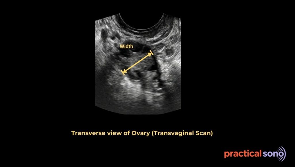
To measure the width, rotate the transducer 90 degrees from the longitudinal position. This produces a transverse (cross-sectional) view of the ovary.
In this view, place the calipers horizontally across the widest part of the ovary. Ensure that the measurement represents the maximum width.
Calculating Ovarian Volume
After entering the length, depth, and width, the ultrasound machine automatically calculates the ovarian volume using its built-in formula.
Formula for Ovarian Volume:
The standard formula used to calculate ovarian volume is
Ovarian volume = Length × Width × Depth × 0.523
This formula is based on the assumption that the ovary has an ellipsoid shape.
However, it is not necessary to memorize this formula. Modern ultrasound machines already have this calculation built into their system. Once the sonographer measures the length, width, and anterior-posterior depth, the machine automatically calculates the ovarian volume.
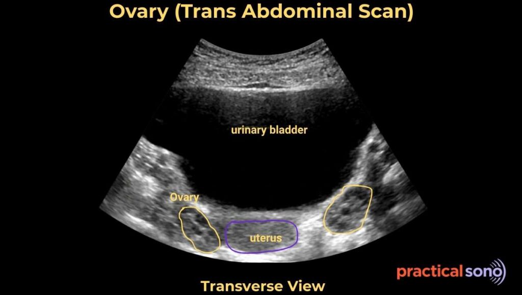
Example on Transabdominal Ultrasound
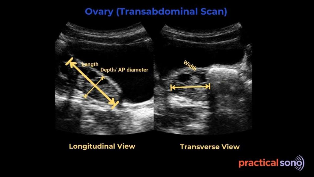
In a transabdominal ultrasound image, the ovary is first evaluated in the longitudinal view.
In this view:
- The length of the ovary is measured from the upper to the lower margin.
- The anteroposterior diameter (depth) is measured from the anterior margin to the posterior margin.
Next, the probe is rotated to obtain the transverse view. In this view, the width of the ovary is measured across its widest dimension.
These three measurements are then used by the ultrasound machine to automatically calculate the ovarian volume.
Clinical Importance of Measuring Ovarian Volume

Measuring ovarian volume has several important clinical applications.
First, it helps in identifying Polycystic Ovary Syndrome (PCOS). Enlarged ovarian volume is one of the diagnostic features used when evaluating patients for this condition.
Second, it can help detect serious conditions such as ovarian torsion. In torsion, the affected ovary often becomes enlarged due to edema and impaired blood flow.
Third, ovarian volume can assist in the screening of postmenopausal ovarian tumors. Comparing the size of both ovaries may help detect abnormal enlargement that requires further evaluation.
Finally, ovarian volume is considered a reliable marker of ovarian reserve. It helps clinicians estimate how a patient might respond to fertility treatments such as in vitro fertilization (IVF).
For these reasons, measuring ovarian volume is an essential part of a complete pelvic ultrasound examination.
Normal Ovarian Volume at Different Ages
Ovarian size changes significantly throughout life. Age and hormonal status strongly influence the volume and appearance of the ovary on ultrasound.
Ovarian Volume During the Reproductive Years
During the childbearing years, the ovaries reach their maximum size.
Typical ovarian measurements are:
- Length: about 3 to 5 cm
- Width: about 1.5 to 3 cm
- Thickness: about 0.6 to 1.5 cm
The normal ovarian volume usually ranges between 3 and 10 mL.
Ovarian Volume in Newborns and Children
In newborns, the ovary may contain small cysts. This happens because of stimulation from maternal hormones during pregnancy.
After birth, these hormones gradually decrease. As a result, the ovary becomes much smaller, and the minimum ovarian volume is usually around 1 to 2 mL.
Before puberty, the ovary often appears solid on ultrasound. This is because the primordial follicles are extremely small and contain very little surrounding fluid. Therefore, follicles are usually not clearly visible at this stage.
As a girl approaches menarche (her first menstrual period), hormonal activity increases. The ovarian volume then rises rapidly, and follicles become more visible on ultrasound.
Ovarian Volume After Menopause
After menopause, the ovaries become significantly smaller.
The follicles are no longer visible, which makes the ovary more difficult to identify on ultrasound.
In postmenopausal women, ovarian volume usually ranges from 1.2 to 5.8 mL, and in most cases it is less than 4 mL.
The ovarian size typically becomes stable about 10 years after the final menstrual period.
Causes of Increased Ovarian Volume
An increase in ovarian volume can occur for many reasons. Some causes are normal and temporary, while others may indicate disease or even a medical emergency.
These causes can be broadly divided into physiological, benign, emergency, and malignant conditions.
Physiological Causes
Physiological causes are related to the normal hormonal changes of the menstrual cycle.
A dominant follicle or corpus luteum can temporarily enlarge the ovary. A single functional cyst may reach 2.5 to 3 cm in size. This can temporarily double the ovarian volume during that cycle.
Pregnancy is another normal cause of ovarian enlargement. Hormonal stimulation during pregnancy increases ovarian volume, and the corpus luteum of pregnancy is usually present. In such cases, ovarian volume may increase to around 14 mL.
Ovarian hyperstimulation syndrome is another physiological condition. It is often seen during in vitro fertilization (IVF) treatment. Multiple follicles develop at the same time, which can lead to massive enlargement of both ovaries.
Benign Causes
Several benign conditions can also increase ovarian volume.
Polycystic Ovary Syndrome (PCOS) is one of the most common causes. It is typically defined as an ovarian volume greater than 10 mL, especially when no dominant follicle is present.
An endometrioma, also known as a chocolate cyst, can also enlarge the ovary. It is a fluid-filled cyst that may appear almost solid on ultrasound. A characteristic feature is the presence of ground-glass echoes within the cyst.
Benign ovarian tumors may also increase ovarian size.
A dermoid cyst, also called a mature cystic teratoma, may contain fat, hair, or even teeth. These cysts can grow quite large.
Other benign tumors include serous cystadenomas and mucinous cystadenomas. These are thin-walled cystic tumors that can grow to a very large size and significantly increase ovarian volume.
Emergency Causes
Some causes of ovarian enlargement require immediate medical attention.
Ovarian torsion is a surgical emergency. The ovary becomes massively enlarged due to venous and lymphatic obstruction. In many cases, the ovarian volume may increase to more than 15 to 20 mL.
Another emergency condition is a tubo-ovarian abscess, which occurs as part of pelvic inflammatory disease (PID). In this condition, the ovary becomes enlarged and inflamed. It often appears attached to the fallopian tube, forming a complex inflammatory mass.
Malignant Causes
Malignant conditions can also cause an increase in ovarian volume.
Primary ovarian cancer may present as a solid mass or a complex cystic lesion, often resulting in significant ovarian enlargement.Metastatic tumors can also involve the ovaries. One well-known example is a Krukenberg tumor, which usually causes bilateral ovarian enlargement. These tumors most commonly originate from gastrointestinal or breast cancers.
Key Takeaway
Understanding the causes of ovarian enlargement helps clinicians determine whether the change in size is normal, concerning, or a medical emergency.
Measuring ovarian volume is the most accurate way to assess ovarian size on ultrasound. It provides more reliable information than a single measurement and plays an essential role in diagnosing PCOS, detecting ovarian torsion, evaluating ovarian reserve, and assessing postmenopausal ovaries.