Renal Cysts are common finding while doing abdominal ultrasound, especially in person over age 60.
What is renal cyst?
It is a fluid-filled sac found in a kidney with otherwise normal parenchyma. It does not communicate with the collecting system.
It appears round and anechoic lesion in ultrasound. If you see such lesion during scan, ask your self these questions:
- Is it simple renal cyst?
- Or, there are any features that makes it a complex cyst?
Simple cyst are asymptomatic and does not need any treatment. They are harmless incidental finding.
But complex cyst can be infection, hematoma, or malignancy. So you must differentiate simple cyst with complex one.
After reading this tutorial, you will
- know the ultrasound features of simple renal cyst
- know the ultrasound features of complex renal cyst
- understand the standard measurement technique for renal cyst
- know the common differential diagnosis
- know how to differentiate simple vs complex cyst (infection, hemorrhage, and malignancy)
- know how to report your finding
- learn about Bosniak classification system
Ultrasound Features of Simple Renal cyst
To call a cyst simple, it should have these ultrasound features:
- Shape: Round or oval . It appears oval in all scanning planes.
- Wall and margin: Thin, smooth, imperceptible. It should not have any mural nodules
- Internal echoes: Anechoic (completely black).
- Strong posterior acoustic enhancement: Because the cyst contains clear fluid, it exhibits acoustic enhancement (brightness behind the cyst), which confirms its fluid-filled nature.
- No Internal Structures: It is not septated (no internal walls) and contains no internal echoes, calcification or solid components.
- No Connection: It should not be connected to the renal collecting system.
- No Blood Flow: On color Doppler, there should be no blood flow associated with the cyst.
Because many complex or malignant conditions can mimic a cyst, a simple cyst is considered a diagnosis of exclusion; all other suspicious features must be ruled out before it is labeled simple.
If all these sonographic criteria are met, and cyst looks simple, no further evaluation or follow-up of the cyst is required
Complex renal cyst have following ultrasound features:
- Thick wall
- Mural nodularity
- Presence of Internal Echoes
- Septation
- Solid Componenent
- Calcification
- Presence of vascularity on color Doppler
These features are described in later in this article. So, keep reading.
Location of simple Renal Cyst
Simple cysts can be found in various parts of the kidney, including:
- The poles (the most common location).
- The cortex or medulla.
- The center of the parenchyma.
- Rarely, they can be exophytic, meaning they bulge out from the surface of the kidney.
Parapelvic Cyst:
It is considered a type of simple renal cyst. They are benign, fluid-filled lesions originating in the renal parenchyma that protrude into the renal sinus, frequently causing pain or obstruction.
It often mimic hydronephrosis on imaging. It is distinguished by a lack of communication with the renal calyces and a distinct separation from the renal pelvis. These cysts are characterized by their location in the hilar region and a potential for causing obstructive symptoms due to their proximity to the renal pelvis..
Number of renal cyst:
Bilateral Presentation: It is very common to have a few simple cysts in both kidneys simultaneously, especially as people age.
Unilateral Presentation: In rarer cases, multiple cysts might be found in only one kidney, while the other remains completely clear.
Localized Cluster: Sometimes, several cysts appear grouped together in one specific section of a kidney rather than being spread out.
Essentially, while simple cysts are often single, having a “handful” of them in various locations is normal and usually does not indicate a more serious genetic condition like Polycystic Kidney Disease (PKD)
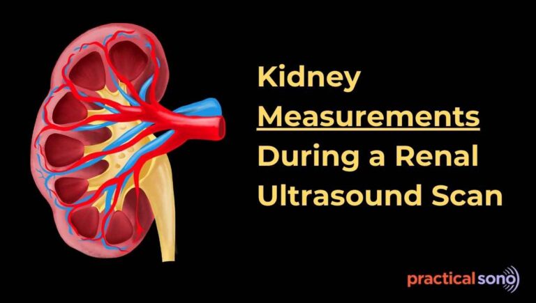
Size of Renal cyst : Measurement technique
How to measure it? To get an accurate size, the cyst is measured in three dimensions:
- Obtain image of cyst in transverse and longitudinal planes.
- Visualize maximum dimension in each planes.
- Once the full cyst is seen freeze the image. Wall of cyst should be fully visualized and not obscured.
- In longitudinal plane, length and height is measured. Length is longest dimension in long axis. Height is anterior posterior measurement.
- Rotate the 90 degree to get transverse plane. Measure width. It is horizontal measurement.
- Calipers at the inner walls of the cyst, NOT the outerwall ensuring the measurement represents the fluid-filled cavity and not the surrounding tissue.
- Place them perpendicular to wall.
Interpretation:
Most simple cysts are small, often ranging from the size approx. 1–4 cm. While many remain stable, some can double in size over a 10-year period.
In some cohorts, an average annual growth of about 3.8% or roughly 3–5 mm per year has been observed. A cyst is generally considered “large” once it exceeds 5 cm in diameter.
Clinical Clue
Usually asymptomatic, discovered incidentally on ultrasound. Large cysts may cause dull flank pain, abdominal mass, or rarely hematuria. No risk of malignant transformation in a true simple cyst.
The occurrence of simple renal cysts is highly correlated with age:
- Children: They are very rare, occurring in less than 0.5% of the pediatric population.
- Adults: Incidence increases significantly over time. More than 10% of adults aged 50 or older have them, and this number rises to over 70% in adults older than 70 years.
Pathophysiology
The exact cause of simple renal cysts is unknown, but they are thought to be acquired rather than inherited.
One theory suggests they form when small, fluid-filled sacs form on the tubules of the nephron, the kidney’s filtering unit.
These sacs may detach and develop into simple cysts. Do not communicate with the collecting system.
Differential Diagnosis
Several conditions can mimic a simple cyst on an ultrasound and must be differentiated:
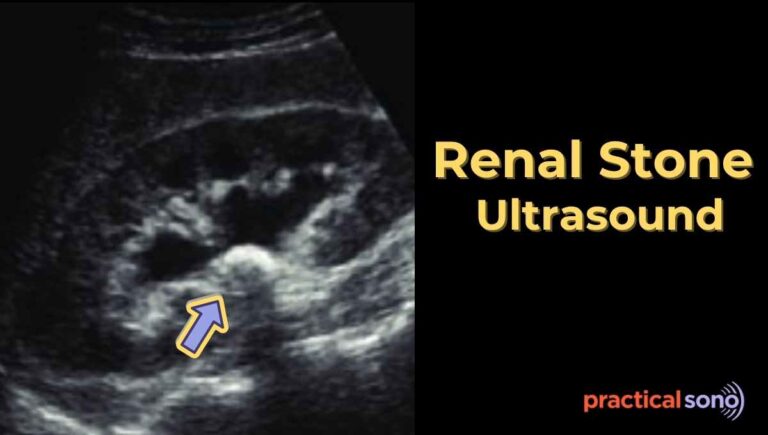
- Hydrocalyx: present with stone obstructing calyx of kidney causing hydrocalyx or caliectasis.
- Pseudoaneurysm: A vascular lesion that can look like a cyst but will show circular blood flow when using color Doppler.
- Calyceal Diverticulum: not differentiated in ultrasound. a diverticulum will fill with contra st on an IVP because it communicates with the collecting system.
- Early Polycystic Kidney Disease: In its early stages, a hereditary disease may present with only one or two cysts. It can be manifestation of other cystic disease in child.
Reporting Simple Ultrasound (Sample)
Finding: A well-defined, round, thin-walled, anechoic lesion measuring 3.2 × 2.5 cm in the upper-pole of the right kidney, showing posterior acoustic enhancement. No septations, solid components, or internal vascularity.
Impression: Features are consistent with a simple renal cyst.
Diagnosis and Management
No treatment needed for simple cysts unless symptomatic or very large (>5 cm with compression effect) Prognosis of a solitary simple cyst is generally good.
In Children:
A simple cyst requires more caution as it may be the first manifestation of a more serious condition, such as polycystic kidney disease.
Consequently, children require a detailed family history, clinical examination, and at least one follow-up assessment. Child with first time diagnosis. It is diagnosis of exclusion.
Detailed medical and family history should be taken. Through clinical examination and at least one follow up assessment should be done. Usually, in children, it does not need further evaluation with contrast enhanced US, MRI or CT imaging.
In Adults:
If a cyst is confirmed to be simple, no further evaluation, follow-up, or imaging (such as CT, MRI, or contrast-enhanced ultrasound) is typically not necessary.
Simple vs Complex Renal Cyst
Complex renal cysts are cystic lesions in the kidney that do not meet the strict criteria of a simple renal cyst.
It is very important to identify a complex cyst because it can sometimes harbor or develop into cancer, such as cystic renal cell carcinoma.
Now let us look at the characteristics of a complex cyst.
- A complex cyst may show mural nodularity.
- The wall may be clearly visible, irregular, or thickened.
- Internal echoes may be present inside the cyst.
- Septations may be seen within the cyst.
- A solid component may be present.
- Calcification may also be seen.
- On color Doppler, vascularity may be detected.
When you see these characteristics, a CT scan is usually required to confirm the diagnosis.
Now, let us look at the characteristics of a complex cyst in detail:
Wall Characteristics
A simple cyst has a thin and smooth wall, whereas, a complex cyst often has a thick wall or a mural nodule.
The presence of a soft tissue mass or mural nodularity, which means small bumps on the wall, is an important feature of a complex cyst.
Irregular or thickened walls are suspicious for necrotic tumors.
Rupture of Renal Cyst
Rupture of a renal cyst can cause acute flank pain. On ultrasound, fluid may be seen in the perinephric space adjacent to the cyst. The kidney may appear fixed.
Internal Echoes
Complex cyst have internal echoes. These echoes may be caused by fluid debris levels. They are either fresh blood clot, infection, or tumor.
About 6% of cysts are complicated by hemorrhage. Infection of a cyst may occur through hematogenous spread, vesico-ureteric reflux, or as a complication of cyst puncture or surgical manipulation.
Let’s look at some example of complex renal cyst with internal echoes:
#1 Hemorrhagic Cyst
Unlike simple cysts, hemorrhagic cysts contain internal echoes or fluid debris levels. These echoes are usually caused by blood or a fresh blood clot.
One major challenge is distinguishing a blood clot from a solid tumor.
Blood clot vs Solid tumor:
- A fresh clot may appear as a soft tissue mass within the cyst. However, the clot is usually mobile. It may move when the patient changes position, such as from a supine position to a side lying position.
- Color Doppler is very helpful in this situation. A hemorrhagic clot shows no blood flow on color Doppler, whereas, solid tumor or necrotic mass often shows vascularity.
Cysts that are suspected to be hemorrhagic on ultrasound may be followed with serial ultrasound.
#2 Infected Cyst
An infected cyst may show a thickened cyst wall. A debris fluid level or gas fluid level may also be present.
Other ultrasound features include a thick wall and internal echoes.
Echogenic perinephric fat may be seen around the cyst. The kidney may appear fixed. There may also be probe tenderness during the scan.
Patients often have clinical symptoms such as fever with leukocytosis. Infected cysts usually require aspiration and drainage for diagnosis and treatment.
#3 Hydatid Cyst
Hydatid cysts may sometimes appear as a unilocular cystic mass. In such cases, they may be difficult to distinguish from a simple cyst.
However, hydatid cysts are usually multilocular cystic masses.
They often contain multiple daughter cysts, which is a characteristic feature.
The cyst wall may also become calcified due to the death of the parasite.
Calcification in Renal Cysts
Calcification can be seen in some renal cysts. It may appears as
- fine and linear,
- thick and amorphous,
- or as milk of calcium.
Fine, linear calcification in the wall or septa without a solid component or thick septa is often not significant.
However, thick and amorphous calcification is more concerning and may require biopsy to rule out malignancy.
Benign vs malignant:
Thin wall or septal calcification usually suggests a complicated cyst rather than malignancy. If all other ultrasound features of a benign cyst are present and there is no soft tissue mass enhancement on CT, the cyst is usually benign.
However, thick, irregular, or amorphous calcification is more suspicious.
In such cases, further evaluation with CT is recommended.
Milk of Calcium:
Milk of calcium is a special type of calcification that can occur inside renal cysts.
It is generally considered a sign of a benign condition.
The most important feature of milk of calcium is a fluid to fluid level inside the cyst.
This happens because calcium particles are suspended in the cystic fluid and settle at the bottom due to gravity.
Another key feature is positional shifting. Because these particles are mobile, the fluid level changes when the patient changes position during the scan.
Septations in Renal Cysts
Septations are internal walls inside a cyst.
Thin septa that are less than 1 millimeter thick and show no blood flow on Doppler are usually benign.
However, thick septa greater than 1 millimeter or septa that show blood flow on Doppler may suggest a tumor.
Sometimes it is important to differentiate a septated cyst from two adjacent cysts.
Occasionally, two nearby cysts may appear like one large septated cyst.
In some cases, it may not be possible to clearly differentiate them.
If the septa are thin or barely visible, smooth, and attach to the cyst wall without thickened areas, the cyst is usually benign.
For example, you may see focal septal thickening greater than 1 millimeter.
There may also be calcification within the septum.
Tumor in a Cystic Lesion
A tumor may show thick septa. Blood flow may be seen within the septa on Doppler.
Internal echoes and a solid area may also be present within the cyst.
Color Doppler may show flow within the thickened septa or the solid component.
Necrotic Tumor: A necrotic tumor may show thick and irregular walls.
Increased blood flow may be seen within the wall on Doppler.
Complex cyst vs solid lesion: Sometimes a cyst may contain internal echoes.
Posterior acoustic shadowing may suggest that it is a cyst.
However, a solid appearing area may create confusion between a tumor and a blood clot.
In such cases, color Doppler should be used. If blood flow is seen within the mass, it suggests a tumor.
Bosniak classification system
The assessment of the malignant potential of renal cysts based on their complexity on imaging forms the basis of the Bosniak classification system.
This classification was introduced in 1986 by Bosniak.
The role of ultrasound in the Bosniak classification system is not fully established.
This is because the Bosniak criteria are mainly based on contrast enhanced cross sectional imaging.
When a cystic renal mass does not meet the criteria of a simple cyst, further imaging is usually required.
In most cases, a renal mass protocol CT scan or MRI is recommended.
An exception may be cysts that show only thin and few septa. These are usually classified as Bosniak category 2 cysts.
Key Takeaway
Simple renal cyst are common incidental finding during routine abdomen ultrasound. It is common in old age.
Simple renal cyst can be easily diagnose due to it unique ultrasound features:
- Round or oval shape
- Thin wall
- Anechoic lumen
- Posterior acoustic shadow
- No internal echoes
- No vascularity on color doppler
When you see such lesion on kidney, you need to differentiate it from the complex cyst, which might be infection, hemorrhage, or even malignancy.