As a beginners, when you scan bowel, all you see is gas or fluid filled tubular structure. Unlike other abdominal organ like liver and kidney, it is very difficult to identify bowel on ultrasound.
It can be very confusing and intimidating. What you need is systematic approach to bowel ultrasound.
After reading this tutorial, you will
- know the most common bowel pathology that you’ll frequently see
- get the ultimate checklist to assess the bowel
- understand the difference between small bowel and large bowel
- know the standard measurement technique for wall thickness and bowel diameter
- know how to use the checklist to diagnose common bowel pathology
Important: You cannot diagnose every bowel condition using ultrasound. Only about 85 to 90 percent of clinically important bowel diseases can be detected this way. However, this represents a major chunk of pathology. To master bowel ultrasound, you need to focus on these common conditions.
Many other rare conditions are diagnosed using CT, MRI, endoscopy, or histopathology. Remember, ultrasound alone cannot reliably detect all bowel diseases, and you do not need to know every single one.
If you understand the common bowel pathologies and know how to diagnose them using the checklist provided in this article, you are good to go.
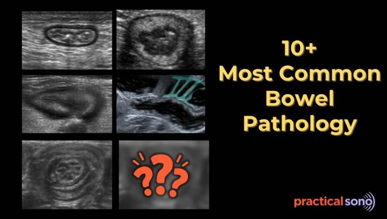
List of most common bowel pathologies seen on Ultrasound (based on location)
RIGHT SIDED PATHOLOGY
- Acute appendicitis
- Mesenteric Lymphadenitis
- Crohn’s disease
- Intussusception
- Ileocecal TB
LEFT SIDED PATHOLOGY
- Diverticulitis
- Epiploic Appendagitis
- Ischemic Colitis
VARIABEL LOCATION
- Bowel obstruction
- Infectious Colitis
- Enteritis
- Ileus
- Colon Cancer
- Gastric Carcinoma
PracticalSono Tips:
Around 70–80% bowel pathologies that can be diagnosed on ultrasound are:
- Appendicitis
- Obstruction
- Diverticulitis
- Infectious/inflammatory colitis
- Enteritis
- Ileus
Another 10–15% will include:
- Crohn’s disease
- Malignancy
- Ischemic
- Intussusception
- Abdominal tuberculosis
Remaining 5–10% will be rare or non-visualizable conditions.
Ultimate Checklist to Assess Bowel on Ultrasound
- Clinical clue ( Sign and symptoms, Investigation report)
- Is it large bowel or small bowel pathology?
- Wall thickness measurement
- Is it symmetrical or asymmetrical?
- Bowel diameter measurement
- Gut layers/ Gut signature
- Is it intact or disrupted?
- Peristalsis and luminal content
- Is it increased or decreased?
- Check the compressibility
- Is it compressible or not?
- Check vascularity
- Is it increased or absent?
- Look for associated finding (Secondary Sign)
- Inflammation of surrounding mesenteric fat (fat stranding)
- Fluid around lesion
- Free fluid in peritoneal cavity
- Air within wall
- Lymph node status
Now, we’ll explain each checklist in detail. Also, we’ll tell you how it help you diagnose specific bowel pathology.
#1 Clinical Clues
Before you start scanning the bowel, you must first look for clinical clues that suggest the probable pathology.
First, ask about the patient’s history and review investigation report.
This will guide you toward the correct differential diagnosis.
For example, an older patient with weight loss, constipation, and blood in the stool should raise suspicion for colon cancer. In such cases, you should focus on the colon and carefully look for both direct and indirect ultrasound signs.
Always know your patient’s history before starting the exam. This helps you know what to look for and where to look.
Next, ask the patient where it hurts the most.
If the patient can clearly point to the most painful area, there is a high chance that the pathology is located there.
Carefully examine that area first. After that, scan the rest of the abdomen.
The location of pain can also give you clues about the possible disease affecting the patient. Certain bowel diseases commonly occur in specific areas of the abdomen.
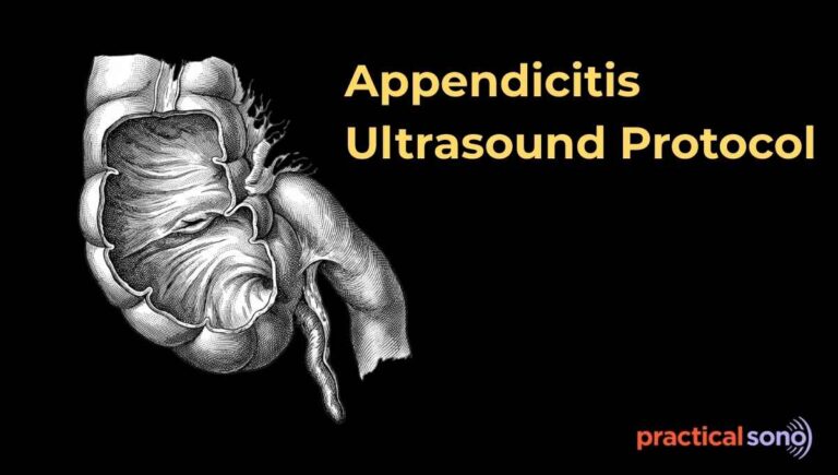
For example, in appendicitis the pain is usually located in the right lower quadrant of the abdomen. Appendicitis is also most commonly found in this area.
So when a patient has right lower quadrant pain, you should carefully examine that region and look for signs of an inflamed appendix.
Intussusception is common in children. The most common type is ileocolic intussusception. It is usually seen in the right upper quadrant of the abdomen, near the right kidney and the liver. So when you suspect intussusception in a child, you should carefully scan that area.
Knowing these patterns helps you focus your search during the ultrasound examination.
Of course, you must still scan the entire bowel in a systematic way. However, when the patient’s symptoms and investigation findings suggest a specific disease in a certain part of the bowel, you should spend more time examining that region to confirm your diagnosis.
#2 Small Bowel or Large Bowel
Another important step during bowel ultrasound is to decide whether you are looking at the small bowel or the large bowel. Let us understand how to differentiate them on ultrasound.
Large Bowel:
Location is an important clue. The ascending colon, descending colon, and rectum are fixed in the retroperitoneum.
This means they usually stay in the same position. However, the transverse colon is not fixed and can move more freely.
So, scan in these areas to find the large bowel.
Haustration: The colon can also be identified by the presence of haustrations.
These are the sac-like segments of the colon. Haustrations are easier to see when the colon is filled with fluid. When the bowel is collapsed or empty, these features become harder to identify.
Small Bowel:
Location is also helpful when identifying the small bowel. Most small bowel loops are not fixed. They move freely within the abdomen, similar to the transverse colon.
Folds of Kerckring: The small bowel is characterized by the folds of Kerckring. These folds are seen within the bowel wall. You will usually see more of these folds in the jejunum than in the ileum.
Up to this point, using clinical clues, you should have a differential diagnosis in mind and know which part of the bowel might be affected. You also know how to differentiate between the small and large bowel on an ultrasound.
Next, we will explore the differences between a normal and an abnormal bowel, as well as the checklist you need to tick off to confirm your diagnosis.
PracticalSono Tips:
The mass effects – Remember, a normal and collapsed bowel is often not well visualized on ultrasound. However, when there is pathology in the bowel, it usually becomes easier to see because it creates a mass effect.
A thickened bowel wall, especially when it is associated with abnormalities in the surrounding soft tissues, produces a clear mass effect. This can be easily recognized on ultrasound.
In addition, a thickened bowel segment often contains less gas, which improves sonographic visualization.
If you suspect any abnormality or see mass effect on any bowel segment, carefully analyze that segment of bowel. Keep the following points in your mind while evaluating it.
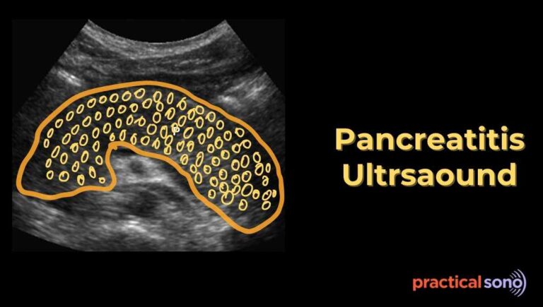
#3 Bowel Wall Thickness
Always start by looking at the bowel segment carefully. Compare it with the adjacent bowel loop. Ask yourself if it looks thicker than the nearby bowel.
If it appears thicker, freeze the image and measure it to confirm.
Measure the wall thickness.
Measure it from the outer hyperechoic serosal surface to the inner echogenic mucosal luminal interface.
Do not include the contents inside the lumen. Do not measure from serosa to opposite serosa. Measure only a single wall thickness.
Apply minimal probe pressure while measuring.
Interpretation:
- In a distended small bowel, the wall thickness should be 3 mm or less.
- In a non distended small bowel, the wall thickness should be 4 mm or less. Any value beyond these limits should raise suspicion for pathology.
Next, determine whether the bowel wall thickening is symmetrical or asymmetrical.
This is one of the most useful signs when differentiating bowel diseases.
Symmetrical thickening:
It means the bowel wall is evenly thick all around the lumen. This pattern usually suggests infection, inflammatory bowel disease, or ischemia.
Asymmetrical thickening:
It means one side of the bowel wall is thicker than the other. The bowel lumen may also appear narrowed. This pattern is highly suspicious for malignancy or a localized tumor.
#4 Gut layer/stratification: Is it intact or lost?
After identifying bowel wall thickening, the next important question is: Do you still see the normal layered appearance of the bowel wall?
A normal bowel shows five alternating bright (echogenic) and dark (hypoechoic) layers, called the gut signature.
Normal Bowel Wall Layers (from lumen to serosa)
- Echogenic interface (lumen–mucosa interface)
This is the bright line facing the lumen. It represents the interface between the bowel contents and the mucosa. - Hypoechoic mucosa
This is a thin, dark layer just beneath the luminal interface. It can thicken early in inflammatory conditions. - Echogenic submucosa
This is a bright, relatively thick layer. It becomes more prominent when there is edema or hemorrhage. - Hypoechoic muscularis propria
This is a dark layer responsible for bowel peristalsis. It can thicken in chronic inflammation or obstructive conditions. - Echogenic serosa
This is the thin outer bright line. It represents the serosal surface of the bowel.
Interpretation:
- Preserved stratification (Layered Pattern Visible
This means the layers are still distinguishable.
Suggests:
• Infectious colitis
• Enteritis
• Early inflammatory bowel disease
• Mild ischemia
• Edema
PracticalSono Tips: Preserved stratification = usually benign / inflammatory.
- Loss of stratification (Gut Signature Lost)
The wall appears homogeneously hypoechoic or heterogeneous.
Individual layers cannot be distinguished.
Suggests:
• Advanced inflammation
• Severe ischemia
• Malignancy
#5 Bowel Diameter Assessment
Check whether the bowel is dilated or collapsed.
For the small bowel, the diameter varies depending on meals but is usually less than 2.5 to 3 cm.
A diameter greater than 3 cm may suggest an obstruction.
For the large bowel, normal diameters are different.
The colon is usually normal up to 6 cm, and the cecum may measure up to 9 cm. Any dilatation beyond these values suggests obstruction or ileus.
#6 Bowel Peristalsis
Observe the dynamic movement of the bowel loops in real time.
Check for the presence, absence, or exaggeration of bowel motion.
Normal peristalsis appears as smooth, wave-like contractions that push the contents forward.
Hyperperistalsis, or increased motion, can be seen in obstruction or infection.
Hypoperistalsis, or absent motion, may indicate ileus or ischemia.
Any abnormality in peristalsis, whether increased, decreased, or absent, is important.
A “to-and-fro” motion is a classic sign of small bowel obstruction.
#7 Check vascularity
Normal bowel shows only mild submucosal blood flow. Minimal or no vascular signal is usually detected.
Increased vascularity, or hyperemia, suggests inflammation, such as in Crohn’s disease.
Absent vascularity may indicate ischemia or necrosis.
In inflammatory bowel disease, semiquantitative grading systems like the Limberg score can help assess the severity of bowel wall vascularity.
#8 Compressibility
Assess the bowel wall by applying gentle pressure with the probe.
Normal bowel is easily compressible and deforms without resistance.
Non-compressible loops suggest inflammation, fibrosis, mass, or obstruction.
Stiff or decreased compressibility indicates possible tumor infiltration or chronic inflammatory changes.
#9 Luminal Content
Observe the contents inside the bowel.
Fluid appears anechoic (dark), while gas appears as bright echogenic foci with reverberation artifacts.
Debris or abnormal content may suggest infection or obstruction.
Dilated loops filled with fluid, gas, or debris can indicate obstruction.
The “small bowel feces sign” refers to particulate matter seen within a dilated small bowel loop, which is a key sign of obstruction.
#10 Associated Findings in Bowel Pathology
Fat Stranding
On ultrasound, fat stranding appears as bright, non-compressible tissue surrounding the bowel.
This is a hallmark of active, full-thickness inflammation.
The fat reacts to inflammatory signals coming through the bowel wall.
Fluid Around Lesion
Small pockets of dark, anechoic fluid seen next to a thickened bowel segment.
This suggests localized leakage or severe inflammation. It can precede a contained perforation or early abscess formation.
Ascites
Fluid that collects generally in the abdomen, often in the pelvis or around the liver.
While it can result from liver or heart disease, in bowel pathology it usually indicates severe systemic inflammation, protein loss, or generalized peritonitis.
Air Within the Wall
Appears as bright echogenic spots or streaks within the bowel wall layers, sometimes with “dirty” shadowing.
This is a medical emergency. It indicates bowel wall ischemia or severe infection that allows gas to enter the wall.
Lymph Nodes
Enlarged, oval or round, dark structures in the mesentery near the affected bowel.
- Inflammatory: Common in conditions like Crohn’s disease.
- Malignant: Very round, large lymph nodes over 10 mm that lose their fatty center may suggest lymphoma or adenocarcinoma.
Key Takeaway
Bowel ultrasound becomes easy and less intimidating when you use a systematic approach.
Fortunately, the diseased segment of the bowel is usually easy to see on an ultrasound, which is a big relief.
All you have to do is look for the direct and indirect signs that lead to the final diagnosis.