A horseshoe kidney is the most common type of renal fusion anomaly. It is often rotated and located abnormally low. This make it difficult to diagnose.
After reading this tutorial, you will
- know what is Horseshoe kidney and its anatomy
- understand its complication
- recognize ultrasound features of Horseshoe kidney
- know the common pearls and pitfalls.
Horseshoe Kidney occurs in approximately 1 in every 400 births.
In this condition, the two kidneys are joined together, usually at their lower poles. The bridge connecting the kidneys is called the isthmus.
Normally, the kidneys develop as two separate bean-shaped organs on either side of the spine.
In a horseshoe kidney, the kidneys fuse together and form a U-shaped structure that crosses the midline of the body.
Most people with a horseshoe kidney have no symptoms and may never know they have the condition.
It is often discovered incidentally during an ultrasound or CT scan performed for another reason.
Understanding the anatomy and ultrasound appearance of a horseshoe kidney is important because it is associated with several complications, including hydronephrosis, kidney stones, and urinary tract infections.
What Is a Horseshoe Kidney?
A horseshoe kidney forms during fetal development when the two kidneys fuse together before they complete their normal ascent into the abdomen.
In most cases, the lower poles of the kidneys fuse together.
The connecting bridge is called the isthmus.
The isthmus may contain:
- Normal functioning renal tissue
- Fibrous tissue without kidney function
Because the isthmus becomes trapped beneath the inferior mesenteric artery during development, the kidneys cannot ascend to their normal position.
As a result, horseshoe kidneys are located lower in the abdomen than normal kidneys.
Anatomy of a Horseshoe Kidney
In a normal person:
- The kidneys are separate.
- The renal pelvis faces medially.
- The lower poles are farther apart than the upper poles.
In a horseshoe kidney:
- The kidneys are fused.
- The lower poles are closer together.
- The upper poles are more lateral.
- The renal pelvis often faces anteriorly rather than medially.
- The fused kidney crosses the midline.
Each kidney still has:
- Its own renal pelvis
- Its own ureter
However, the orientation is often abnormal because of malrotation.
Blood Supply
The blood supply of a horseshoe kidney can be complex.
The main blood supply comes from the abdominal aorta.
Additional arteries may arise from:
- Inferior mesenteric artery
- Common iliac artery
- Internal iliac artery
- External iliac artery
Multiple renal arteries are common.
This is important to remember when planning surgery or other interventions.
Common Complications of Horseshoe Kidney
Most patients have no symptoms.
However, abnormal anatomy can interfere with normal urine drainage and increase the risk of complications.
1. Kidney Stones
Kidney stones are one of the most common complications.
Why does this happen?
The ureters must pass over the isthmus and often follow an abnormal course.
This can slow urine drainage.
Slow drainage leads to urine stasis, which increases the risk of stone formation.
2. Hydronephrosis
Hydronephrosis is also common.
Many patients develop obstruction at the ureteropelvic junction (UPJ).
The ureter may become stretched or compressed as it crosses the fused bridge of tissue.
This obstructs urine flow and causes dilation of the collecting system.
3. Urinary Tract Infection
Urinary tract infections occur more frequently in patients with horseshoe kidneys.
Why?
Urine that drains slowly allows bacteria to multiply more easily.
Repeated infections may occur if urinary stasis is present.
4. Increased Risk of Trauma
Horseshoe kidneys lie lower in the abdomen than normal kidneys.
They also sit directly in front of the spine.
Because of this position, they are more vulnerable to injury during blunt abdominal trauma, such as:
- Motor vehicle accidents
- Falls
- Crush injuries
Ultrasound Findings of Horseshoe Kidney
Now let’s look at the sonographic appearance.
Kidneys Located Lower Than Normal:
The kidneys are usually positioned lower in the abdomen or pelvis.
This occurs because the fused kidneys cannot complete their normal upward migration during fetal development.
When scanning, a kidney that appears unusually low should raise suspicion for a horseshoe kidney.
Lower Poles Point Toward the Midline:
One of the most helpful clues is the orientation of the kidneys.
Normal kidneys have lower poles that point away from each other.
In a horseshoe kidney, the lower poles point toward the spine and the midline.
This creates an inverted V appearance.
Difficulty Visualizing the Inferior Pole:
On a routine longitudinal flank scan, the lower pole may appear to disappear toward the midline.
This happens because the kidneys curve inward toward the fused isthmus.
If you can see the upper pole clearly but struggle to visualize the lower pole, consider the possibility of a horseshoe kidney.
Presence of an Isthmus:
The most important finding is the identification of the isthmus.
The isthmus appears as a bridge connecting both kidneys across the midline.
It is usually located:
- Anterior to the abdominal aorta
- Anterior to the inferior vena cava
- Just below the origin of the inferior mesenteric artery
Parts of Isthmus
- Functional renal tissue
- Fibrous isthmus
Functional Renal Tissue
Most isthmuses contain functioning renal tissue.
In these cases, the isthmus appears:
- Similar in echogenicity to the renal cortex
- Continuous with both kidneys
Fibrous Isthmus
Sometimes the isthmus contains only fibrous tissue.
In this situation, it appears as:
- Thin
- Hyperechoic
- Difficult to visualize
Transverse Scan Appearance:
During a transverse sweep through the mid-abdomen, the isthmus may appear as a soft tissue structure crossing the midline.
It often looks draped over:
- The abdominal aorta
- The inferior vena cava
- The vertebral column
Malrotated Renal Pelvis:
The renal pelvis and ureteropelvic junction often face:
- Anteriorly
- Laterally
Instead of facing medially as in a normal kidney.
This abnormal orientation is called malrotation.
Complications to Look for During Ultrasound
Whenever you identify a horseshoe kidney, always search carefully for associated complications.
Hydronephrosis:
Look for:
- Dilated renal pelvis
- Dilated calyces
- Evidence of UPJ obstruction
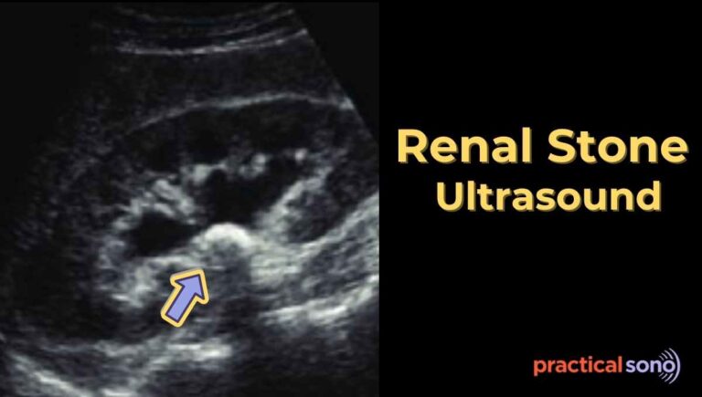
Kidney Stones:
Look for:
- Echogenic foci
- Posterior acoustic shadowing
- Twinkle artifact on color Doppler
Renal Cysts:
Renal cysts may occur within a horseshoe kidney.
Because of distorted anatomy, small cysts can be easy to miss.
Renal Masses:
Tumors can also occur within a horseshoe kidney.
Carefully evaluate the entire renal parenchyma because abnormal anatomy may obscure lesions.
Pearls
Visualizing the isthmus can sometimes be difficult.
The most common problem is bowel gas in the midline.
Helpful techniques include:
- Using graded compression
- Scanning through the liver window
- Scanning through the spleen window
- Performing a slow transverse sweep across the midline
These methods often improve visualization of the isthmus.
Common Pitfalls
The isthmus can be mistaken for other structures.
It may resemble:
- Pre-aortic lymph nodes
- A soft tissue mass
- An abdominal aortic aneurysm
Always follow the tissue carefully and determine whether it connects both kidneys.
Finding continuity with the kidneys confirms the diagnosis.
Example Ultrasound Report
Findings:
Both kidneys are located lower than expected within the abdomen. The lower poles are fused across the midline by an isthmus of renal tissue located anterior to the abdominal aorta. Renal pelves demonstrate mild anterior orientation. No hydronephrosis or renal calculi are identified.
Impression:
Findings are consistent with a horseshoe kidney. No associated complication is identified on this examination.
Key Takeaway
A horseshoe kidney is the most common renal fusion anomaly and occurs when the lower poles of both kidneys fuse during fetal development.
The key ultrasound findings include:
- Low-lying kidneys
- Lower poles directed toward the midline
- Presence of an isthmus crossing the midline
- Malrotated renal pelvis
- Inverted V appearance of the kidneys
Whenever a horseshoe kidney is identified, always evaluate for:
- Hydronephrosis
- Kidney stones
- Urinary tract infection
- Renal masses
Recognizing these characteristic ultrasound features will help you confidently diagnose a horseshoe kidney and identify associated complications.