Note not every bright echo with shadowing in kidney is renal stone. There are lots of renal stone mimickers.
And, you should know them to avoid misinterpretation and misdiagnosis.
After reading this short article, you will:
- know common pathologies that mimic renal stone on ultrasound.
- learn why these mimicker appears
- and, how to differentiate them from true renal stone (most important)
Why Do Stone Mimickers Occur on Ultrasound?
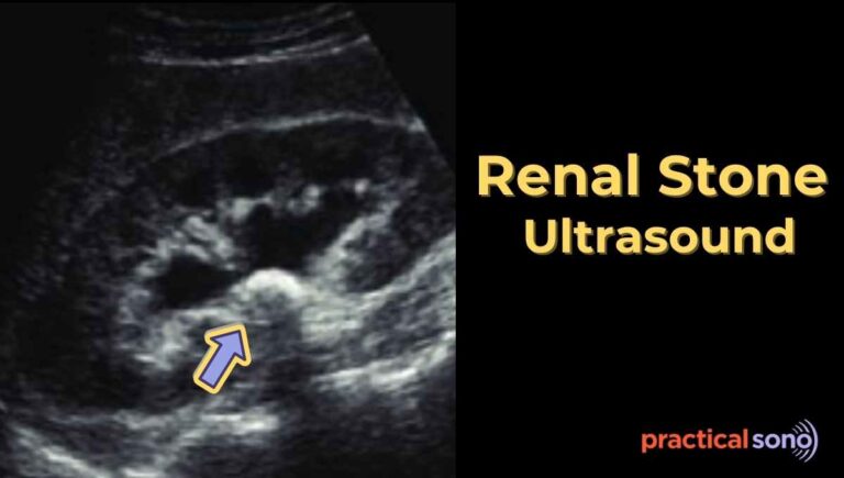
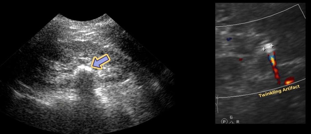
A true kidney stone usually appears as a
- bright (echogenic) focus
- shows posterior acoustic shadowing
- and, have a twinkle artifact on color Doppler.
However, other conditions can also show bright echoes, shadowing, or twinkle artifact. That is why careful scanning and clinical correlation are very important during renal scan.
Common Renal Stone Mimickers on Ultrasound
1. Vascular Calcifications (Renal Artery Calcification)
Calcified renal arteries, especially arcuate or segmental arteries, can appear as bright echoes inside the kidney.
They mimic stones by producing bright echoes, sometimes shadowing, and twinkle artifact.
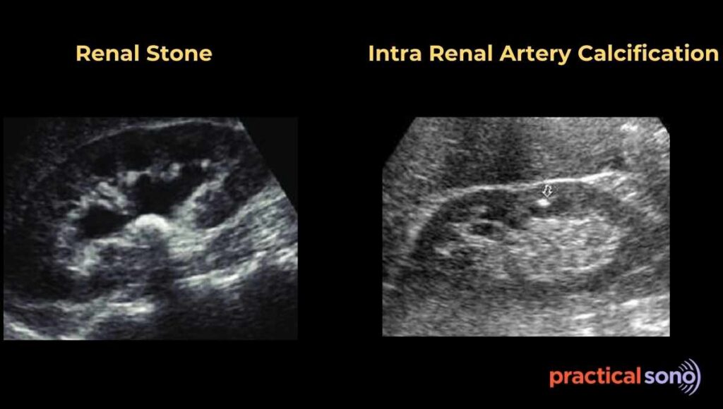
To differentiate:
Renal artery calcification is located along blood vessels, often regularly spaced, and color doppler shows blood flow within it. If Doppler shows flow, it is not a stone.
Remember, true renal stone are often present in urine collecting system such calyx, pelvis, and ureter. That’s why you need to look at these region for true renal stone.
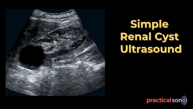
2. Milk of Calcium Cyst
This cyst contains calcium particles suspended in fluid. It can occur in a simple renal cyst or a calyceal diverticulum.
On ultrasound, it looks like a fluid-filled lesion with layered bright material that may show shadowing. The material shifts with patient movement.
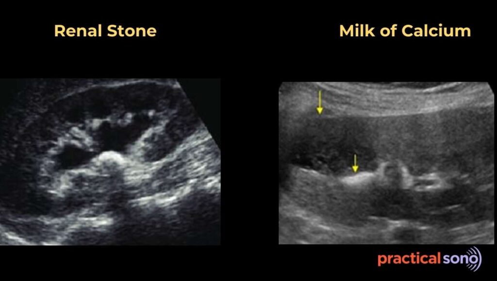
If the bright echoes moves when the patient changes position, think milk of calcium, not a stone.
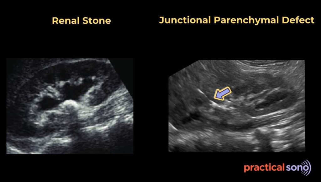
3. Junctional Parenchymal Defect
This is a small congenital fusion defect of the kidney.
It appears as an echogenic line or triangular defect, usually in the upper pole. It follows the kidney contour and does not have the typical shadowing of a stone.
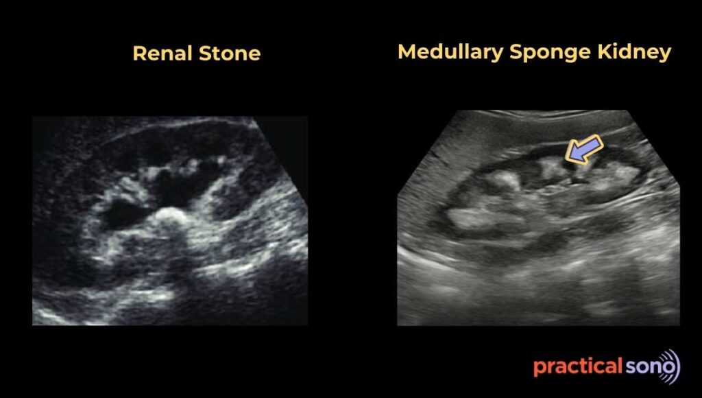
4. Medullary Sponge Kidney
It is a congenital condition with dilated collecting tubules in the kidney medulla. These tubules may calcify.
Ultrasound shows clusters of bright medullary echoes with multiple small calcifications. Both kidneys are often affected. Unlike a single stone, there are multiple small foci.
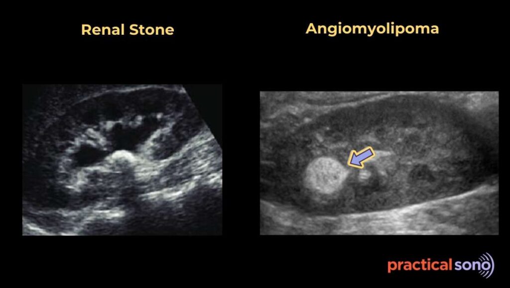
5. Angiomyolipoma (AML)
AML is a benign kidney tumor made of fat, vessels, and muscle.
It looks bright on ultrasound and can mimic a small stone. It appears as a solid mass, usually in the cortex, and often does not show clean shadowing. Fat-poor AML can be tricky.
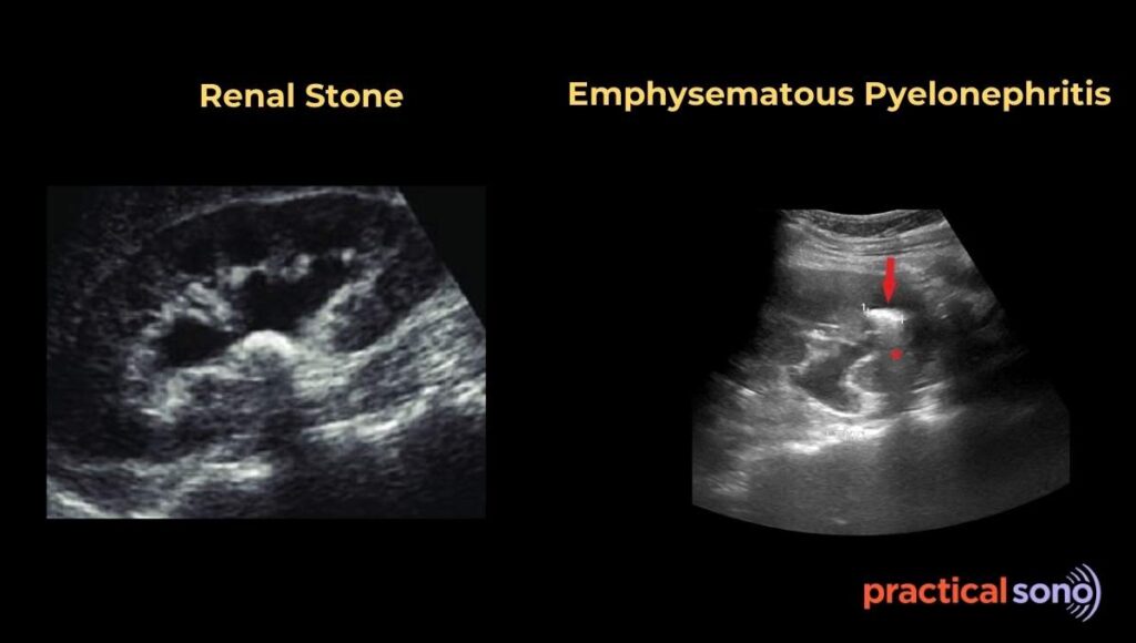
6. Intrarenal Gas
Gas inside the kidney creates very bright echoes with dirty shadowing.
It occurs in severe infections such as emphysematous pyelonephritis. Ultrasound shows reverberation artifact and dirty shadowing. Clinical signs of infection help differentiate it from a stone.
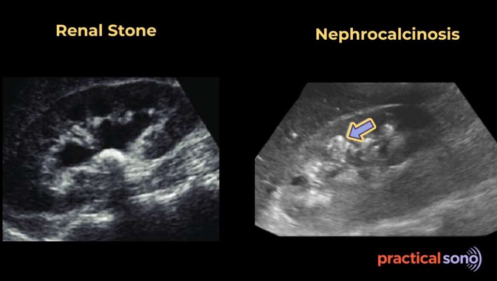
7. Nephrocalcinosis
Nephrocalcinosis is a condition where too much calcium builds up inside cortex and medulla of the kidneys.
Kidney Stones are lumps that form in the open spaces where urine flows. (Urine Collecting System)
Nephrocalcinosis is tiny calcium deposits scattered throughout renal parenchyma. (Cortex and Medulla)
Types of Nephrocalcinosis
- Medullary (98% of cases)
- Cortical (Rare)
Normally, medulla appear anechoic triangular area.
In Nephrocalcinosis, these pyramids become very bright white (hyperechoic) instead of dark.
In early stages, you might only see a bright white ring around the edge of the pyramids.
In severe cases, the entire middle of the kidney looks solid white.
In Nephrocalcinosis, these pyramids become very bright white (hyperechoic) instead of dark.
In early stages, you might only see a bright white ring around the edge of the pyramids.
In severe cases, the entire middle of the kidney looks solid white.
Key Takeaway
Before calling a bright focus a stone, ask yourself:
- Does it have clean posterior shadowing?
- Is there twinkle artifact?
- What is the exact location? Is it in urine collecting system? Or, within parenchyma.
- Does the patient’s sign and symptoms support renal stone?
Remember, not every bright echo in the kidney is a stone. Careful scanning, doppler evaluation, and clinical correlation are the keys to accurate diagnosis.