Not all hydro nephrosis is pathological. And, ureteric stone is not only the cause of hydro nephrosis.
After reading this article, you will
- know how to diagnose hydro nephrosis and its grading
- learn common causes
- and common pitfalls and pearls.
Normal Sonographic Anatomy of Kidney
To understand the hydro nephrosis and its grading, first, let’s quickly review the normal sonographic anatomy of kidney.
Renal capsule: This is renal capsule, it appears as bright, thin, hyperechoic line that perfectly demarcates the kidney from the surrounding perirenal fat. Below it is cortex and medulla.
Renal Cortex: Cortex is outer rim of renal parenchyma. This has a fine, uniform, grainy grey appearance. Its brightness is normally isoechoic or hypoechoic compared to a healthy liver or spleen.
Medulla: It appears as series of anechoic triangular or wedge-shaped just inside the cortex. These are renal pyramids. They appears more prominent in children and think adults.
Renal sinus: It is central core. It has renal calyces, renal pelvis and major blood vessel within it. Also, it contains a high concentration of fat. Due to this fat, it appears as hyperechoic area on ultrasound.
Urine collecting system: In normal healthy kidney, the collecting system calyces, renal pelvis, and ureter are typically not visible becauue they are collapsed and hidden within the bright sinus fat. They are visible only when it is dilated due to urine stasis.
Urine collecting system
To understand the grading of hydronephrosis, you need to have clear picture of urine collecting system in your mind.
Urine is formed in nephron which is present in the cortex and medulla.
Multiple nephrons drain their fluid into these larger tubes located within the renal pyramids.
At the very tip of the pyramid, the collecting ducts merge into larger papillary ducts.
These ducts open onto the surface of the renal papilla through small holes. Urine drips through these openings directly into the minor calyx. Typically, there is one minor calyx for every renal pyramid. There are about 8 to 18 minor calyces in each kidney.
Usually, 2 to 3 minor calyces merge to form a major calyx.
The major calyces coalesce into the funnel-shaped renal pelvis. This is the largest, central funnel where urine first collects before entering the ureter.
From renal pelvis, urine flow into ureters. Ureters have peristaltic waves that squeeze urine downward toward the bladder every 10 to 15 seconds.
Ureter have natural anatomical constrictions at ureteropelvic junction, pelvic brim crossing and ureterovescial junction. These are the location where ureter stone gets lodged.
The urinary bladder serves as a expandable storage reservoir. It is lined with transitional epithelium that allows it to stretch as it fills with up to 500 mL of urine.
Ureter does not enter into the bladder in straight line. Instead of going straight through the bladder wall, the ureter travels through it at an angle for about 1 to 2 cm. As the bladder fills with urine and its walls stretch, they naturally press against this “tunneled” section of the ureter.
This pressure squeezes the ureter shut, acting as a one-way valve that prevents urine from flowing backward (refluxing) toward the kidneys.
When you are ready to urinate, the bladder’s detrusor muscle contracts, and the sphincter muscles relax, allowing urine to exit the body through the urethra.
If you’ve understood this pathway of urine flow clearly, it becomes easy to understand the cause of hydronephrosis and its grading.
Ultrasound features of Hydro-nephrosis
There is grading for hydronephrosis which tells how much urine has “backed up” into the kidney’s drainage system. So how you know how much urine has “backed up”.
Why you need it?
- It directly influence the clinical management plan. It tells whether patient need follow up scan or equire urgent surgical intervention to prevent permanent kidney damage. Higher the grades, increased risk of renal function deterioration. Lower the grades are, it is often benign and self limiting.
- Grading following standard system will make sure we’re talking same things while discussing with collegue.
- Also to see if the swelling is stable, improving, or worsening over time,
Most common grading system we follow is that give by society for fetal urology. According to this system, there are four grade of hydronephrosis.
We’ll observe three anatomical features: the renal pelvis, the calyces, and the renal parenchyma
Grade 0 (Normal): There is no visible dilation. The walls of the collecting system are touching each other, and no anechoic fluid is seen in the center.
Grade 1 (Mild): There is slight dilation of the renal pelvis only. The urine appears as a small black slit that barely splits the bright center of the kidney.
Grade 2 (Mild): The renal pelvis is moderately dilated, and a few major calyces (but not all) are now visible as fluid-filled pockets.
Grade 3 (Moderate): Virtually all major and minor calyces are uniformly dilated. The internal borders between the pelvis and calyces are still recognizable, and the renal cortex is still a normal thickness.
Grade 4 (Severe): This is marked by “gross” dilation where the pelvis and calyces appear “ballooned” or merged into a single large fluid sac. The critical indicator for this grade is parenchymal thinning (cortical atrophy), where the filtering tissue of the kidney is visibly squashed by the high pressure.
There are many other grading systems for the assessment of the degree of hydronephrosis have been proposed. Grading system by society for fetal urology (SFU) is commonly used.
Caliectasis
Hydronephrosis is general swelling of entire renal collecting system, both pelvis and calyces. However, if only calcyces is dilated, it is called focal Caliectasis.
Small stone that obstructs a minor or major calyx can cause localized caliectasis.
Other cause include, urinary tract infection, renal fibrosis or stricture or tumor or cyst applying pressure to calyces causing localized back up of urine.
On ultrasound, it appears as anechoic, fluid-filled dilation of one or just few specific calyces, rather than the entire collecting system.
Renal pelvis often remains normal in size or is significantly less dilated than the affected calyx. This is hallmark finding.
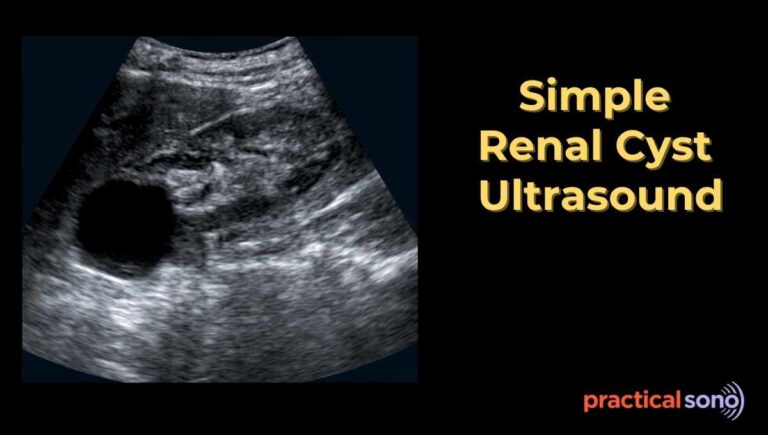
It is often get confused with renal simple cyst.
The shape of focal Caliectasis is often irregular or “branching” pattern. It connects to renal pelvis. It is found within the area of collecting system.
Simple renal cyst is round or oval in shape. It does not communicated with renal pelvis. It has thin and smooth wall. It is never thick. In caliectasis, wall can be thick if infected. Simple renal cyst can be anywhere in the cortex or medulla. It is not limited to the collecting system.
False Negative Case
Sometimes, kidney may appears normal even if there is blockage in renal collecting system.
Visual expansion of the kidney’s collecting system depends on pressure created by the accumulation of urine. If the body is not producing enough urine to build up that pressure, the structures will not stretch or “dilate” enough to be seen on an ultrasound.
This happens in severe dehydration or in renal failure.
When a patient is severely dehydrated, The kidneys produce very little urine, and there is not enough volume to fill the area behind a blockage and force it to expand.
In dehydrated patient, give 500ml of IV fluid bolus and rescan later for the hydronephrosis.
In acute kidney injury or advanced renal failure, the kidneys lose their ability to filter blood effectively. If the kidneys are barely functioning, they may stop producing urine altogether. Without urine flow, the collecting system remains collapsed even if a stone or tumor is blocking the path.
False Positive Case
A false positive means the scan shows black fluid areas, but there is no blockage.
Para pelvic cyst: Cysts are simple fluid sacs. The key differentiator is connectivity. Hydronephrosis branches and connects back to the renal pelvis; cysts are isolated “bubbles” that don’t lead anywhere.
Prominent Renal Pyramids:
In thin adult, or children, renal pyramids appear very dark and can be mistaken for dilated calyces. However, they are arranged in a perfect geometric ring and never “merge” into the center.
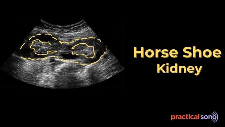
Extra renal pelvis: In some people, the renal pelvis sits outside the kidney rather than inside. It can look very large and “swollen,” but if the calyces inside the kidney are not dilated, it’s a harmless anatomical variant.
Over hydration: A very full bladder creates back-pressure. If you see swelling, ask the patient to go pee and rescan. If the swelling disappears, it was just a full bladder or temporary reflux.
Common cause of hydronephrosis:
These are the common cause of hydronephrosis:
Obstructive
- Intrinsic (Within collecting system)
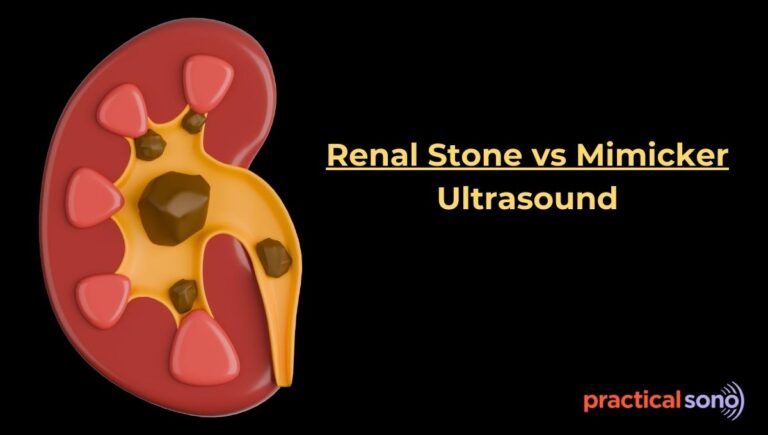
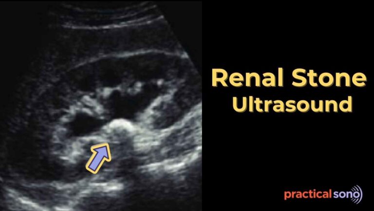
- Renal or ureteral stones (common)
- Blood clots
- Neurogenic bladder
- Posterior urethral valve
- Ureterocele
- Extrinsic (Outside collecting system)
- Prostate hypertrophy
- Pelvic tumors (large fibroids, ovarian carcinoma)
- Pregnancy (seen in 2nd trimester)
- The right ureter is frequently affected than left ureter
Non-obstructive
- Vesicoureteral reflux
- Urinary tract infection
- High flow states ((Diabetes insipidus, Psychogenic polydipsia)
- Distended bladder
Key Takeaway
Whenever you see hydro nephrosis, do these things:
- Is it physiological or pathological? Rule out any false positive or false negative condition.
- Find out the cause of hydronephrosis. If there is unilateral hydronephrosis, trace the ureter. Try to find the cause, most of the time you’ll see the ureteric stone.
- Then, assess the grade of hydronephrosis.
- See how it has compressed the cortex of kidney. High pressure within the collecting system can cause atrophy of cortex. Measure the cortical thickness. Normal thickness is more than 10mm. Once the cortex thickness is less than 7mm, it is sign of chronic pressure. Once this tissue is scarred or lost, the damage is often irreversible leading chronic kidney disease and renal failure. Also, Stagnant urine acts as a breeding ground for bacteria, significantly increasing the risk of Urinary Tract Infections (UTIs) and pyelonephritis.
- Most studies suggest that if a complete obstruction is corrected within 2 weeks, the kidney can often regain nearly normal function.
- If severe hydronephrosis persists beyond 6 to 8 weeks, the likelihood of permanent damage increases significantly.